Anyway, my love for the show stemmed from my need to make, paint, build and mould anything and everything I could. I wasn't an artistic protege (far from it, that was my sister's territory), but I loved it all the same.
So when I got the chance to build a giant re-creation of the human cell with a class of wonderful six-year olds from Wroughton Infant School in Norfolk, I obviously jumped at it. This is the end result:
The children did a fabulous job of making the cell itself (I'll give the details below) and then I filled it with an array organelles. I think it's a great teaching tool, and while I'll mention the names of each of the organelles and a bit about them below, this can obviously be tailored up or down depending on your age group.
So here's how...
The cell
For the structure itself, you need a giant balloon. I bought these on Amazon (6 for a tenner), but I'm sure there are other outlets. This was hung up at the school and then plastered in mod rock (the stuff they use for plaster casts.) You want to put a few layers on this to make it sturdy.
Once it's dried, you've got to get the balloon out (much to the entertainment of the children - see below!). I'm sure you can work out how - a pair of scissors on the end of a stick worked for us. We were expected a pretty big bang, but were rather disappointed!
All that's left now is to cut it into a bowl shape and paint it. We also covered it in tissue before painting it, so make the surface smoother.
Now comes the fun part: the stuff inside...
The "brain" of the cell. All DNA is stored here, and all messages are sent and received from this central control station. The nucleus is actually made up of a central nucleolus, surrounding nucleoplasm and an envelope (to hold it all together!). For our nucleolus, we painted a football black and cut it in half. The nucleoplasm was painted yellow and the envelope black. Getting the football to hold to the mod rock took quite a lot of glue... Normal PVC glue probably isn't going to stand you in good stead for this project - we used a glue gun and an adhesive glue spray.
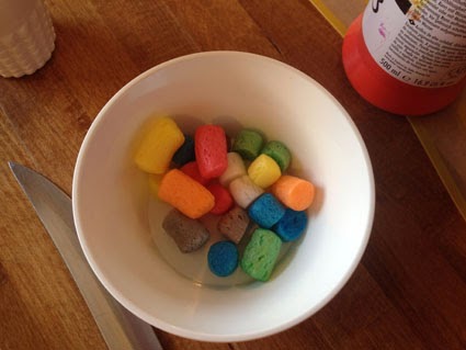
The factory and quality control centre of the cell. Once the nucleus receives a signal, it sends out its own in return, in the form of RNA. This RNA is converted into protein by the ribosomes - those little wotsit like things around the nucleus. These ribosomes are also found on the surface of some of the Endoplasmic Reticulum. I used PlayMais for these.
Once you have your protein, it's sent into the Endoplasmic Reticulum where it's folded into the right shape and checked for errors. The Endoplasmic Reticulum surrounds the nucleus, and looks like long snakes folded back in each other. I used foam for this, which was bought on the market for 20 quid (but we only used about a quarter of it, so it's not too expensive). This required super strength adhesive glue spray, in order to get the foam to bend around the nucleus. Enter dad and boyfriend for cutting the foam and using the adhesive glue...(let's not gender stereotype, it was actually just really cold outside and they were my lovely volunteers).
The Golgi Apparatus
More foam! This organelle sits after the Endoplasmic Reticulum and modifies the proteins that come to it by adding extra bits (such as sugars). It's made of a stack of this long membrane sacks, which you can see in the picture above.
The Mitochondria
These guys are our source of energy - they make ATP, which we need. It's like our food. They have an extra layer of membrane on the inside, so I've made them out of two colours of thin foam, stuck together.
The Proteasome & Lysosome
These are the dustbins of the cell. They churn up anything we don't want - like proteins that have either folded into the wrong shape, or have already finished their job. The proteasome is a long cylinder, where protein is fed in one end and gets cut into pieces while going through.
The Lysosome is a spherical vesicle that contains enzymes that can break down all kinds of biological molecules. For our model, the children painted make up foam pads, and then I cut some to represent the proteasome. I then used pipe cleaners to represent the protein being fed in and coming out of the proteasome. (There were a load of make up pads on offer in M&S, so that's what I used! Bottom line - be thrifty.)
The Lysosome isn't on its own, there are other spherical vesicles called the endosomes. I've used round gems to represent these. Get yourself to Hobbycraft.
Vesicles are the taxi cabs of the cell: they get stuff where it needs to be, which includes moving between organelles and even in and out of the cell itself. I thought the tear drop shaped make up pads were a nice way of representing vesicle budding, so the children painted these and we put them moving from the Endoplasmic Reticulum and the Golgi. I also used them to represent endocytosis (moving into the cell), and lined the vesicle with a pipe cleaner to make it look like the cell membrane was folding in.
Those all important proteins that sit on the surface of our cells and make sure messages are received and transmitted. There are different types, but I've included Receptor Tyrosine Kinases (RTKs) and G-protein coupled receptors (GPCRs). RTKs are half on the outside of the cell (to receive signals) and half on the inside (to send them on). They also have to find their partner before they can pass their signal onto the inside of the cell, so I've put some on by themselves, and some with their partner. Oh, and yes, they're made of make up sponges again - those things are amazing.
The GPCRs are a different kind of receptor - they don't need to find a partner, but as you can see, they take up a lot more room! They span the cell membrane a total of seven times. For these, I punched holes in the cell surface (actually my mum did, I can't be trusted with those sorts of things...) and fed through pipe cleaners represent the different parts of the receptor as it goes in and out of the membrane.
The Cytoskeleton
Our cells can't just hold their shape, they need a scaffold. Our scaffold is called a cytoskeleton, and it's made up of long microtubules that run through the cell. We used thread, with buttons on the end, to stick across the model.
Virus
OK...so this isn't actually part of the cell, but the weird bobbly ball thing was too good a prop to miss up on. It's the perfect shape for a virus, which often have these bumpy coats to them. Looks like our cell has been infected!
I think the end result is pretty cool, and it was actually a lot of fun to put together. You could get the class to make their own, learning about the different parts as they go along, or you could use one as a teaching tool.
Plus, these little guys are amazing:
You can see this model at the London Science Museum tonight, along with some amazing workshops, at the Lates event!


No comments:
Post a Comment