Pages

Tuesday, 15 July 2014

The thesis hole

It's been a while since I've posted anything, mainly due to the fact that I've been writing my PhD thesis. Which is a lot less fun... 

Halfway through and full of caffeine-induced positivity, here's a few tips on how to survive inside - and ultimately crawl out of - the thesis hole. 

1. Don't set yourself a daily word count. 
Don't do it! The chirpy fresh me thought that was the best idea in the world. But on those days when it appears easier to squeeze blood from a stone than type 200 words on that screen, it's NOT a good idea. You will feel as though you are an absolute failure, even though literally no one cares about this except you (and possibly your supervisor if you're really lucky). 

2. Do have an idea of how long each chapter is going to take. 
While word targets are a bad idea, so is writing your discussion two days before hand-in. Chop in up into chapter-size chunks and that giant impossible book starts to become manageable.

3. Change up the scenery. 
I write up between home and work. Home allows me to eat cereal whenever I want and wear pyjama shorts all day. Work allows me to talk to people other than my 85-year old neighbour. Whatever works for you. 

4. Politely request that people stop asking you how it's going. 
Three to four months is a long time for people to say "finished it yet?!". No. Seriously. 

5. Have some fun. 
It's true that there will always be something for you to do and you will constantly feel guilty for that hour you spend on Facebook/watching the Kardashians/drowning sorrows at the pub. But you will go insane without it! Take a day off, interact with human beings, have a laugh. 

6. Pre-warn your friends and family that you might not be the best company. 
This was a big one for me. Even when you're not writing, your head will be so full of crap you just wrote that your social skills may be *slightly* impaired. It's temporary - you'll be back to being the life and soul when this is all over. 

7. OK some advice for the thesis itself...write the most amazing plan ever. 
I'm not talking about a few bullet points, more like a few thousand words. Breakdown each chapter into headings, subheadings and then bullet points on the paragraphs. It's effort, but it'll be a lifesaver when your struggling (see point 1). 

8. START EARLY! 
Oh god I wish I'd started earlier. There are going to be holes in your work (unless you're perfect, congratulations and go somewhere else). While those holes can be explained easily enough, sometimes those couple of experiments (in those months I didn't leave spare) could tie together the loose pieces nicely. Just a thought.

9. STOP DOING EXPERIMENTS AND START WRITING! 
While point 8 is true if you leave time, it's also true that that one last experiment that ties it all together and makes it all make sense ain't gonna happen. That experiment will always lead to another - that's like the rule of research. So put down the pipette and put those great ideas in your discussion (write them in your plan now!) 

10. Last but not least: it's all temporary. 
You will write it, someone will read it and then you'll get your PhD. It will happen. So remember that when you're at the bottom of the hole in your pyjama shorts talking to your 85-year old neighbour about the weather. 

Just me? 

Friday, 4 April 2014

Is excess protein as bad for you as smoking?

I was getting ready for work the other morning when the other half ran in looking a tad panicked. He's a big gym goer, and therefore a big protein fan, and had just read the headlines that PROTEIN CAUSES CANCER.

Sounds like something the daily mail would say doesn't it? 'Cept this time the claim was from some fancy scientists, published in a fancy journal. The newspapers were going mad with the idea that steak and salad was as detrimental to your health as smoking some cigarettes. Really Science Daily

First off, I'd say those sort of suggestions are a bit irresponsible no? We know there's a link between cigarettes and cancer. Black and white, do one and you massively increase the risk of getting the other. It's taken a lot of hard work to get that fact out across the world, and I don't think the guys that published this paper meant that at all. It lead to the NHS releasing this

The paper everyone is talking about came from Professor Valter Longo's laboratory, at the University of Southern California, and was published in Cell Metabolism. This means it's not open access (BOOOO!) and therefore isn't available for everyone to read, but I'll let you keep Game of Thrones for your bedtime reading and do the hard work for you. 

To start with, the title sounds a little different to "Excess protein causes as much cancer as smoking"...

Low Protein Intake Is Associated with a Major Reduction in IGF-1, Cancer, and Overall Mortality in the 65 and Younger but Not Older Population

Basically: lower protein in your diet reduces your risk of cancer if you're younger than 65.

It's a really interesting paper and has got some great data in it. It suggests possible links between high animal protein intake and cancer prevalence in the 50-65 age group (which is pretty narrow).

The trial was carried out on about 6000 American adults aged 50 and upwards, with a follow-up period of 18 years. They also tested some of their findings from the study population on mice and in cells. 

For the 50-65 year old age group, consuming high levels of animal protein (considered to be 20% or more of their diet) resulted in a 4-fold risk of dying of cancer. But, in the over 65s category, higher protein levels were associated with the opposite: a 60% reduction in the number of cancer deaths when compared to the low protein (10% or less) group. 

From this, they suggest that a reduced protein diet, followed by an increase in protein consumption once you're eligible for a bus pass might be a good way to go. But there need to be more studies. 

They also monitored levels of IGF-1 in their participants and found that high levels of the growth factor were found in high-protein consuming groups, and this was then related to the high cancer prevalence. I.e. Lots of protein = more IGF-1 in our system = higher risk of cancer. 

After this, they presented data looking at the effect of protein consumption on tumour growth. Mice on high and low protein diets were injected with cancer cells, and the rate of tumour growth was monitored. They found that while all mice on high protein diets had developed tumours, this dropped by 10-20% in mice in low protein diets. This suggests that high protein levels help to promote tumour growth. Coming back to the IGF-1 stuff from earlier, they also found that the mice on a low protein diet had significantly lower levels of IGF-1 in their system. Again, less protein = less IGF-1 = lower risk of cancer. 

In mice. In one study. While the data is very interesting and compelling, I would say there's no need to throw those special offer steaks out of the freezer. Unless they've been in there a while... 

It could suggest that a low protein diet could help during cancer treatment. This isn't the first time protein and cancer have been mentioned together: high amounts of meat or animal fats in our diet have also been linked to breast (open access paper analysing current studies) and colon cancer (open access paper).  

It appears to be limited to animal protein though, vegetable protein is fine. 

I'm inclined to stick to the "everything in moderation" mentality. On that note...cake anyone? 


Thursday, 20 March 2014

The #NoMakeUpSelfie: why I'm on board

Is it a little narcissistic? Probably. Have I done one? Check it out below. To be honest, while there has been a lot of cynicism attached to the craze (of which I was a part of at first), if it's raising money for cancer research, who the hell cares?! At the time of writing this, over ONE MILLION POUNDS had been raised for Cancer Research UK! That's freaking unbelievable!!

And to all the men out there with their "Just a bunch of silly girls doing something silly" mantra, how is it any different to Movember?! Sit back down.


I heard a crazy fact the other day: out of your taxes, cancer research receives as much as the cost of a London pint (that's about £4.50 for those of you lucky enough not to know) each year. Now I work in cancer research, and I know that £4.50 doesn't go very bloody far. So as far as I'm concerned, the more money raised, the better. 

And as a side note, I don't think the craze of telling women they look beautiful without make up is a bad thing, is it? We get up, cover our faces in glorified paint and head out to work. While I'm aware I'm less likely to scare babies with my make up on, when you step back and look at what a large proportion of Western women do, it's a bit mad no?! 

Everyone's a winner: save money on beauty products, receive a compliment or two, and most importantly (obviously), raise money for cancer research. 

So text BEAT to 70099 to donate £3 to Cancer Research UK, and let's see if we can make it to two million. 

You can also text...

"CCUK21 £3" to 70070 to donate to Children with Cancer UK
"MOBILE" to 70550 to donate £5 to Macmillan Cancer Support

Or you can go to www.breakthrough.org.uk to donate to Breakthrough Breast Cancer

Wednesday, 5 March 2014

Designer Drugs: Our Night At The Museum

On Wednesday, we ran a workshop at the London Science Museum, for the "Bio-revolution" Lates session. We wanted to introduce people to the ideas of investigating protein structure and using it to design drugs to treat cancer. 

As it's a Lates session, most people have got a beer or a glass of wine in hand, making for a very relaxed and entertaining evening! It was a fantastic chance to showcase our new 3D printed protein structures and drugs, and they went down a treat. It was such an invigorating experience; so many people asking questions and getting involved. It was also the most popular Lates session to date - nearly 7000 people showed up!! 

Although it was an incredible amount of fun, I was absolutely gutted that I didn't get the chance to go off and explore the rest of the museum myself. There was so much going on, and so many researchers there ready to get the public involved in their science. 

As for our session, it started with an animated video made by the very talented Jeroen Claus, explaining how some cancers develop and one of the ways in which researchers attempt to target it. You can watch the video here


After that, people got a chance to play with the 3D proteins themselves. We had big 3D printed HER2 proteins, with 3D printed flexible drugs that (may) fit into the ATP pocket (or binding site). We started by observing that while ATP fits, it falls out pretty easily when you tip the protein upside down. It was just a nice way of visualising the fact that ATP binds loosely, and a drug just needs to fit more tightly in order to compete with it. 

Enter our drugs (from the top of the fingers down to the palm): Staurosporine, ATP (for comparison), Lapatinib and Taxol.


Staurosporine fits really well - in fact, it fits too well. It fits so well that it likes to bind to loads of ATP pockets - not just the ones in HER2. The fact that it's so promiscuous means that a lot of messages that we need to be transmitted are blocked along with the HER2 ones, meaning that our cells die. So it's used in the lab, but is rubbish in the clinic. 

Taxol is massive. It's actually a natural molecule that was isolated from the Yew tree, and it took forever to recreate in the chemistry lab because of its unusual structure. That unusual bulky structure also makes it a poor fit for the ATP pocket. You can shove it in if you try (and believe me, some of our visitors definitely tried) but you get the idea that it wouldn't happen in the body naturally. Taxol is used to treat cancer though, as a chemotherapy agent. It targets the cytoskeleton (check out my last post for more info on that), stabilising the cell structure and preventing cell division. 

And finally, lapatinib: our winner. I think it looks like a longer ATP - and a lot of drugs that target these sorts of proteins are modelled on the structure of ATP, because we know that fits. It extends further into the back of the pocket, and will be interacting with the amino acids inside. Lapatinib is used as a secondary treatment for HER2-positive breast cancer in the US. 


This sort of personalised medicine isn't going to solve everything - drug resistance is still a big problem. But it does offer a more specific treatment with fewer side effects that chemotherapy. You can read more about targeted cancer therapies in far greater detail here

Overall, we had a lot of fun! 3D printed proteins were a great way to explain structure and drug development, and hopefully we'll get to use them a lot more in the future. 

Wednesday, 26 February 2014

How to make a giant re-creation of the human cell (you know you want to)...

When I was a kid, I absolutely LOVED Blue Peter. I even managed to get myself a Blue Peter badge one year (for harassing my entire street for their old christmas cards), which my mum then put into the wash. FYI, it's now a White Peter badge.

Anyway, my love for the show stemmed from my need to make, paint, build and mould anything and everything I could. I wasn't an artistic protege (far from it, that was my sister's territory), but I loved it all the same.

So when I got the chance to build a giant re-creation of the human cell with a class of wonderful six-year olds from Wroughton Infant School in Norfolk, I obviously jumped at it. This is the end result:


The children did a fabulous job of making the cell itself (I'll give the details below) and then I filled it with an array organelles. I think it's a great teaching tool, and while I'll mention the names of each of the organelles and a bit about them below, this can obviously be tailored up or down depending on your age group.

So here's how...

The cell

For the structure itself, you need a giant balloon. I bought these on Amazon (6 for a tenner), but I'm sure there are other outlets. This was hung up at the school and then plastered in mod rock (the stuff they use for plaster casts.) You want to put a few layers on this to make it sturdy.


Once it's dried, you've got to get the balloon out (much to the entertainment of the children - see below!). I'm sure you can work out how - a pair of scissors on the end of a stick worked for us. We were expected a pretty big bang, but were rather disappointed!


All that's left now is to cut it into a bowl shape and paint it. We also covered it in tissue before painting it, so make the surface smoother. 


Now comes the fun part: the stuff inside...


The "brain" of the cell. All DNA is stored here, and all messages are sent and received from this central control station. The nucleus is actually made up of a central nucleolus, surrounding nucleoplasm and an envelope (to hold it all together!). For our nucleolus, we painted a football black and cut it in half. The nucleoplasm was painted yellow and the envelope black. Getting the football to hold to the mod rock took quite a lot of glue... Normal PVC glue probably isn't going to stand you in good stead for this project - we used a glue gun and an adhesive glue spray.


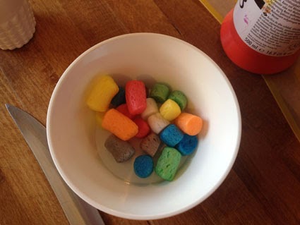
The factory and quality control centre of the cell. Once the nucleus receives a signal, it sends out its own in return, in the form of RNA. This RNA is converted into protein by the ribosomes - those little wotsit like things around the nucleus. These ribosomes are also found on the surface of some of the Endoplasmic Reticulum. I used PlayMais for these.


Once you have your protein, it's sent into the Endoplasmic Reticulum where it's folded into the right shape and checked for errors. The Endoplasmic Reticulum surrounds the nucleus, and looks like long snakes folded back in each other. I used foam for this, which was bought on the market for 20 quid (but we only used about a quarter of it, so it's not too expensive). This required super strength adhesive glue spray, in order to get the foam to bend around the nucleus. Enter dad and boyfriend for cutting the foam and using the adhesive glue...(let's not gender stereotype, it was actually just really cold outside and they were my lovely volunteers).


More foam! This organelle sits after the Endoplasmic Reticulum and modifies the proteins that come to it by adding extra bits (such as sugars). It's made of a stack of this long membrane sacks, which you can see in the picture above.


These guys are our source of energy - they make ATP, which we need. It's like our food. They have an extra layer of membrane on the inside, so I've made them out of two colours of thin foam, stuck together. 


These are the dustbins of the cell. They churn up anything we don't want - like proteins that have either  folded into the wrong shape, or have already finished their job. The proteasome is a long cylinder, where protein is fed in one end and gets cut into pieces while going through. 

The Lysosome is a spherical vesicle that contains enzymes that can break down all kinds of biological molecules. For our model, the children painted make up foam pads, and then I cut some to represent the proteasome. I then used pipe cleaners to represent the protein being fed in and coming out of the proteasome. (There were a load of make up pads on offer in M&S, so that's what I used! Bottom line - be thrifty.) 

The Lysosome isn't on its own, there are other spherical vesicles called the endosomes. I've used round gems to represent these. Get yourself to Hobbycraft. 


Vesicles are the taxi cabs of the cell: they get stuff where it needs to be, which includes moving between organelles and even in and out of the cell itself. I thought the tear drop shaped make up pads were a nice way of representing vesicle budding, so the children painted these and we put them moving from the Endoplasmic Reticulum and the Golgi. I also used them to represent endocytosis (moving into the cell), and lined the vesicle with a pipe cleaner to make it look like the cell membrane was folding in. 


Those all important proteins that sit on the surface of our cells and make sure messages are received and transmitted. There are different types, but I've included Receptor Tyrosine Kinases (RTKs) and G-protein coupled receptors (GPCRs). RTKs are half on the outside of the cell (to receive signals) and half on the inside (to send them on). They also have to find their partner before they can pass their signal onto the inside of the cell, so I've put some on by themselves, and some with their partner. Oh, and yes, they're made of make up sponges again - those things are amazing. 


The GPCRs are a different kind of receptor - they don't need to find a partner, but as you can see, they take up a lot more room! They span the cell membrane a total of seven times. For these, I punched holes in the cell surface (actually my mum did, I can't be trusted with those sorts of things...) and fed through pipe cleaners represent the different parts of the receptor as it goes in and out of the membrane. 


Our cells can't just hold their shape, they need a scaffold. Our scaffold is called a cytoskeleton, and it's made up of long microtubules that run through the cell. We used thread, with buttons on the end, to stick across the model.


Virus

OK...so this isn't actually part of the cell, but the weird bobbly ball thing was too good a prop to miss up on. It's the perfect shape for a virus, which often have these bumpy coats to them. Looks like our cell has been infected!


I think the end result is pretty cool, and it was actually a lot of fun to put together. You could get the class to make their own, learning about the different parts as they go along, or you could use one as a teaching tool.


Plus, these little guys are amazing:


You can see this model at the London Science Museum tonight, along with some amazing workshops, at the Lates event! 


Wednesday, 19 February 2014

Why Structural Biology is awesome.

Structural Biology. I feel it gets a bit of a bad rep: boring, extremely complicated, far removed from actual biology. A strange melting pot of physics, mathematics and chemistry that somehow manages to produce a picture of what something might look like. Plus, the computer programs are really really hard to use.

OK, you're not wrong on the final point - it once took me 2 hours just to work out how to open a program. (Never let pride get the better of you, it's a total waste of time). 

But over the next few posts, I'm going to try and convince you that structural biology is actually pretty cool and (once you've worked out how to open programs...) very accessible. It's not removed from biology either - it's stuck to it like glue. 

I'll start with the basics. Structural biology is just the study of what stuff looks like. How it fits together. But obviously on a microscopic scale. And the end results are pretty beautiful: 


That's the protective antigen portion of the Anthrax toxin. It forms these pores on the surface of cells, allowing the other two pieces of the toxin to enter and do their damage inside the cell. You can find this on the RSCB PDB under the code 1TZO.

Research is carried out on proteins: long chains of amino acids that fold and twist in such a way to produce a huge array of different shapes. And these shapes are incredibly important - the cells in our body don't just know what to do. They need to be told: enter proteins. These guys pass signals from one to the next (aptly named signalling), sending messages between and within cells, telling them what to do. Get bigger; move over there; divide. None of it happens without the constant signalling that occurs via proteins. 

And this is where the shape (or structure of the protein) really comes into play. In order to pass on those signals, they need to be able to bind to each other - like a secret handshake. You want the right protein to meet the right partner - otherwise we'd have signals flying all over the place. Structural biology allows us to look at these unique structures in intricate detail, giving us a better picture of what is actually happening. 

I guess there are two reasons you might want to do this. 

Firstly, scientists love finding out new stuff - it's kind of the point of science. They go out into the unknown, pipette in hand, and come back with loads of new information for us to learn from. 

Secondly, if you don't know what something looks like, how are you supposed to design a drug to target it? When signalling goes wrong, and - for example - we lose the "Stop!" signal while the "Grow!" signal ends up on megaphone, diseases such as cancer can arise. We need drugs that can mute those out-of-control signals, and they therefore have to target whichever protein the signals are coming from. Enter structural biology: this allows us to examine drug-protein interactions on an atomic level, and can help us design more efficient and selective drugs for future treatments. 

If you'd like to learn a bit more about structural biology and drug design, head over to the London Science Museum next Wednesday (26th February from 18:45) for the Lates session. Jeroen Claus and I will be running an interactive workshop on those very things, and there are a tonne of other exciting talks and demonstrations going on all evening. 


Sunday, 16 February 2014

Here goes...

So I've finally got round to starting a blog. It's something I've wanted to do for a long time, but I guess the apprehension of whether anyone is actually interested in my nonsensical ramblings got the better of me.

Until now. 

I'm a structural biology PhD student, so you'll probably find a fair amount of science here. And food. When I'm not pouring this and that into beakers in the lab, I'm probably doing the same thing in my kitchen. With varying results... (that applies to the lab and the kitchen). 

I hope you enjoy... or more to the point, I hope I do...